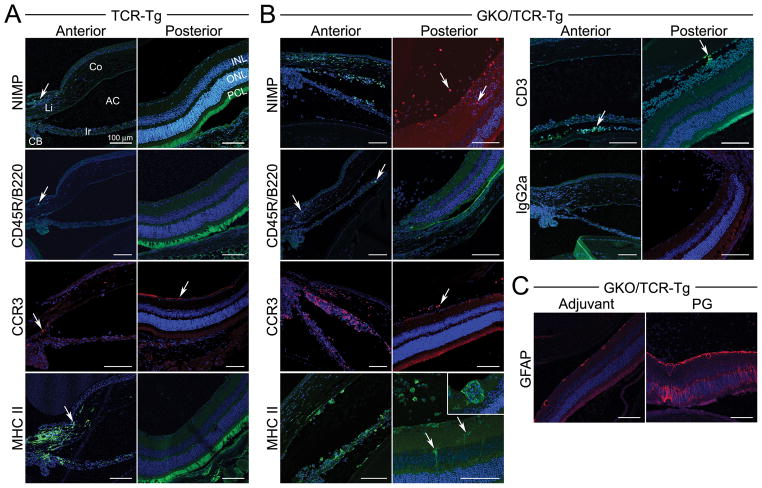
Figure 3. Immunofluorescence staining of uveitic eyes at 3 weeks following immunization.
Eye sections of PG-immunized TCR-Tg (panel A) or GKO/TCR-Tg mice (panel B) that were stained with antibodies for the indicated cell markers (original magnification, 200X): NIMP = neutrophil, CD45R/B220 = B cell, CCR3 = eosinophil (red), MHC Class II (with inset of higher magnification, 400X), CD3 = T cell and IgG2a indicates negative staining in the control antibody group. Panel C) Staining for retinal glial fibrillary acid protein (GFAP) expression within the retina of GKO/TCR-Tg mice in adjuvant controls is increased after immunization, indicating gliosis or astrocyte activation/proliferation. Co = cornea; Ir = iris; CB = ciliary body; AC = anterior chamber; Vi = vitreous; INL, inner nuclear layer, ONL = outer nuclear layer; PCL, = photoreceptor cell layer.

