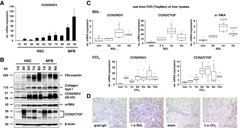
Fig. 1.
CCN3/NOV expression in cultured HSC and in experimental hepatic fibrosis in vivo. a Quantitative RT-PCR (TaqMan) from culture-activated HSC showed significant upregulation of CCN3/NOV, reaching peak levels in fully transdifferentiated MFB. b Western blot analysis of HSC protein lysate, showing increased CCN3/NOV protein expression in culture-activated HSC and MFB that correlated with increased expression of fibronectin, collagen type I, α-SMA and CCN2/CTGF. In this analysis, β-Actin was used as a loading control. c Relative mRNA expression of CCN3/NOV in BDL and chronic CCl4-treated rat livers. Quantitative RT-PCR (TaqMan) showed the relative mRNA levels of CCN3/NOV to increase significantly upon BDL and CCl4 administration. Expression levels were comparable to CCN2/CTGF and α-SMA expression throughout the period of liver fibrogenesis. d CCN3/NOV immunohistochemistry of BDL and CCl4 rat livers reflecting positive staining in mesenchymal cells and proliferative bile duct epithelia along the fibrotic septa of BDL rat livers. The CCl4 model showed positive staining in perisinusoidal areas peripheral to centrilobular hepatic necrosteatosis

