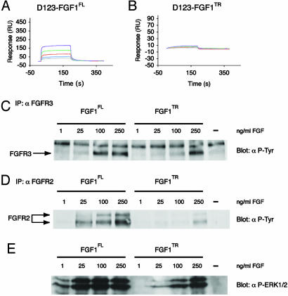
Fig. 2.
The affinity and activation of FGFRs by FGF1 is enhanced by the N terminus. (A and B) Sensorgrams of the full-length versus truncated FGF1 interaction with the D123 isoform of FGFR3c. Analyte concentrations are colored as follows: purple (1.6 μM), green (0.8 μM), red (0.4 μM), blue (0.2μM), and gray (0.1 μM). (C–E) Full-length FGF1 activates FGFR3 and FGFR2 more potently than truncated FGF1 on RCS cells (C–D). Analysis of FGFR3 and FGFR2 phosphorylation. Clarified cell lysates of RCSs starved overnight and treated with the indicated concentration of FGF for 5 min were examined by using protein immunoblot. (E) FGFR3 and FGFR2 immunoblots were probed with a phosphotyrosine-specific antibody. Analysis of ERK1/2 phosphorylation. Clarified cell lysates used in A were probed with an antibody specific for anti-phospho-ERK1/2.

