Fig. 1.
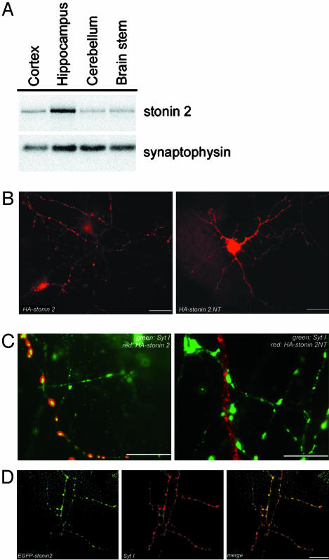
Stonin 2 is enriched in the hippocampus and localizes to axonal vesicle clusters. (A) Western blot of homogenates (50 μg of protein) from different brain regions. (B–D) Localization of stonin 2 in primary neurons. Cortical neurons were transfected with plasmids encoding HA- or enhanced GFP (EGFP)-tagged stonin 2 or a truncation mutant lacking its CT μ-homology domain (stonin 2 NT). Eight days in vitro neurons were analyzed by immunofluorescence microscopy for the distribution of EGFP-stonin 2, HA-tagged stonin 2, HA-tagged stonin 2 NT, and synaptotagmin I. Low (B)- and high (C)-magnification views illustrating the colocalization of HA-stonin 2 but not HA-stonin 2 NT (red; Alexa 594) with synaptotagmin I (green; Alexa 488) at axonal vesicle clusters. (D) Colocalization of EGFP-stonin 2 (green) with synaptotagmin I (red; Alexa 594). (Scale bar, 20 μm.)

