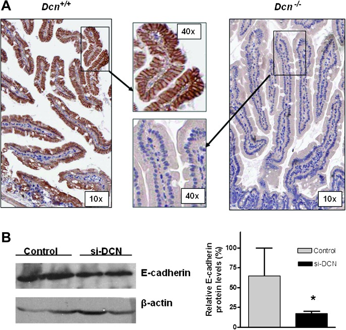
Fig. 1.
Loss of decorin reduced E-cadherin expression in mouse and in vitro. (A) E-cadherin was dramatically reduced in intestinal epithelial cells in Dcn−/− mice by immnunohistochemical staining (anti-E-cadherin antibody dilution 1:100) (magnification ×10 and ×40, respectively). (B) Knockdown decorin expression via decorin siRNA (si-DCN) led to reduced expression of E-cadherin in HEK293 cells compared with the control. The first two lanes stood for that the cell lysated was obtained from duplicated experiments. So did the last two lanes of control. The average levels of E-cadherin were quantified and showed in the right panel. *P < 0.05 compared with the control group.

