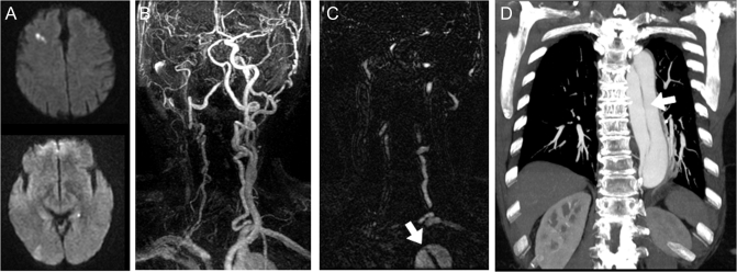
Figure. An 82-year-old woman with acute stroke and painless aortic dissection.
MRI head with (A) diffusion-weighted images showing multiple areas of increased signal intensity (right frontal > right occipital). Contrast-enhanced magnetic resonance angiography of head and neck where (B) right common and internal carotid are not visualized, and (C) a thin black line is identified within the aortic arch on the axial phase of contrasted images thought to represent an intimal flap. (D) Contrast-enhanced chest CT illustrating Stanford type A dissection extending down the abdominal aorta.

