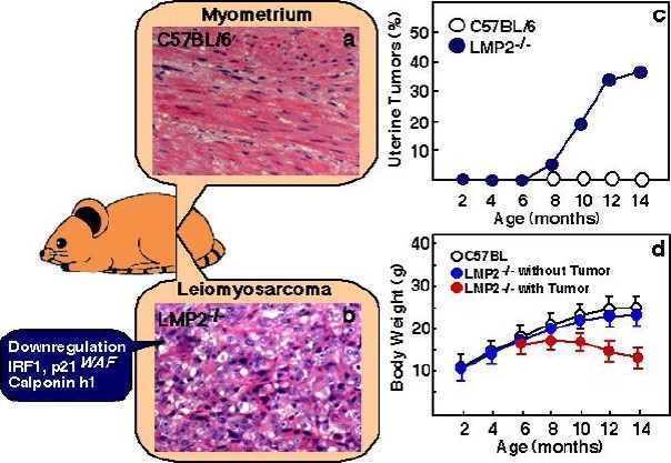
Fig. 1.

Histological findings of myometrium (a) and uterine LMS in LMP2-deficient mice (b). Among the histological findings of uterine LMS in LMP2-deficient mice, a cytoskeleton, which is characteristic of uterine LMS, is observed. (a and b magnification x200) Panel c, in LMP2-deficient females, uterine LMS is observed at 6 months of age. The incidence at age 14 months is as high as 40%. The curve indicating the incidence of mouse uterine LMS is very similar of that indicating the incidence of human uterine LMS, which is observed after menopause. In mice with tumors of the uterus, significant weight loss is observed (d). Thus, a tumor that develops in the uterus is diagnosed as malignant, i.e., uterine LMS. In LMP2-deficient cells, levels of the anti-oncogenic factor IRF-1, p21WAF are significantly reduced. Reduced expression of the calponin h1 transcript, which contributes to cell proliferation and tumorigenesis in myometrium cells, is detected in uterine LMS tissues. The inactivation of such anti-oncogenic factors is considered to transform LMP2-deficient cells into leiomyosarcoma cells.
