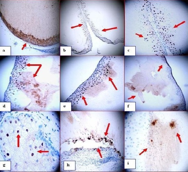
Fig. 2.

a Positive staining with the antibody to Cytokeratin A1/A3 antibody around the wall of the cyst and inside in (red arrow). b through e positive staining with p27 antibody in the wall of the cyst as well as in some spots inside the cyst (red arrows). f. IHC using MMP9 showing some positive staining in the wall of the cyst as well as inside it (red arrows). g. MMP9 (positive in some cells of the sebaceous glands (red arrows) (dark staining). h, MMP9 stain showing how some parts of the cyst are being separated from the adjacent matrix and staining positive for MMP9 on both sites of the gap (red arrow) (dark brown stain). i. Positive staining in some spots inside the cyst using TIMP1 antibody (dark staining, red arrows).
