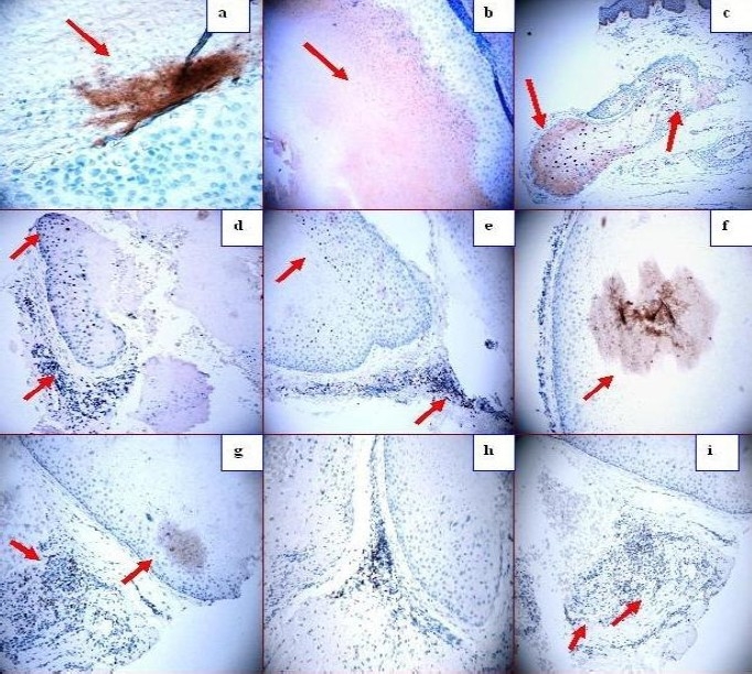
Fig. 3.

a. Zap-70 positive staining in some spots inside the cyst (dark staining; red arrows). b. Positive staining in some spots inside the cyst using TIMP1 antibody (dark staining; red arrows). c, d, and e. p27 positive staining in areas of pilosebaceous units that are apparently “normal” by histology (brown staining; red arrows). p27 stains around other partially damaged cysts, and inside the sebaceous glands (red arrows). f. Positive staining inside the cyst in some patches with alpha 1 anti-trypsin (red arrow). g. BAX positive staining in a patch inside the cyst, and also in the inflammatory infiltrate outside the cyst (dark staining; red arrows). h. BAX positive staining in the inflammatory infiltrate around the cyst (red arrow). i. BCL-10 positive staining in the inflammatory infiltrate around the cyst (red arrow).
