Fig. 2.

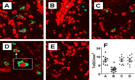
Allergic responses suppress tolerogenic DCs in the nasal mucosa. Mice were treated with the same procedures in Fig.1. Nasal mucosa was taken for immune staining with anti-CD11c and anti-ALDH antibodies. A-D, confocal images show the positive staining of CD11c (in green) and ALDH (in blue). Cells stained in both green and blue were regarded as the tolerogenic DCs and counted (20 fields per mouse) under confocal microscope. Panel D is isotype IgG control. F, scatter dot plots show individual data points in panel A-D. Original magnification: ×200. Insert: ×630.
