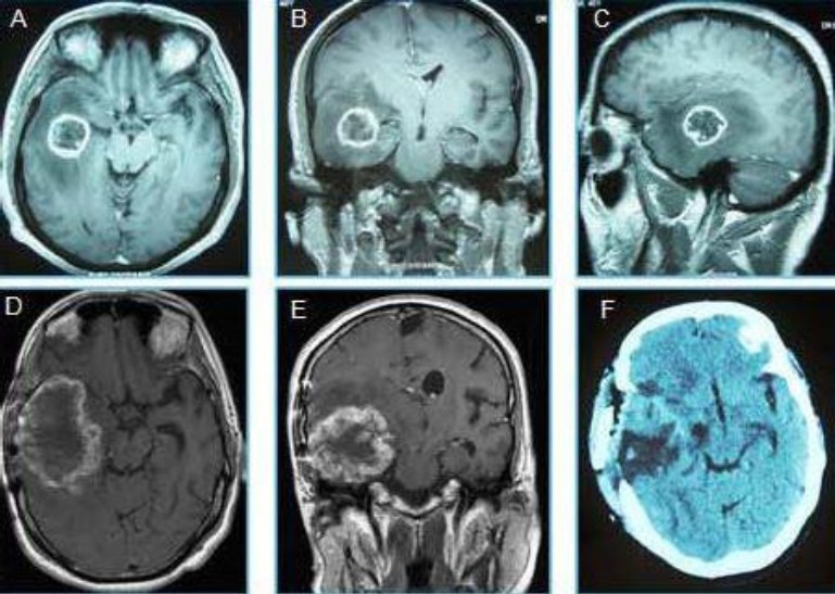
Fig. 1.

Neuroimages of glioblastoma recur after treatment in the same place as gliosarcoma. A: axial, B: Coronary and D: sagittal MRI showed a Right temporal process corresponded to a glioblastoma multiforme before treatment. D: Axial and E: Coronary MRI image of a secondary gliosarcoma, later found at the same location as the previously treated GBM. F: axial CT, performed after the total resection of the gliosarcoma.
