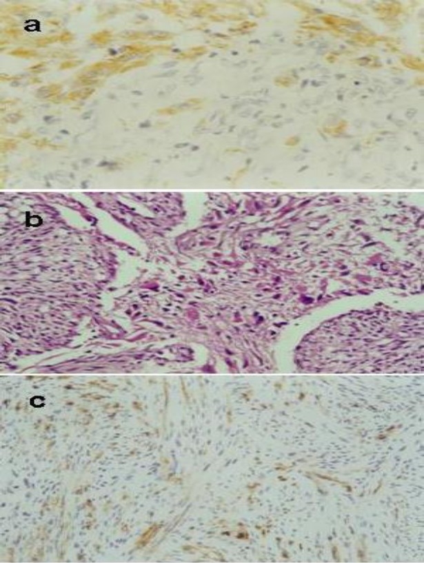
Fig. 2.

Photomicrographs of the tumor. a: The gliomatous component GFAP positive (×100). b: Tumour characterised by a biphasic tissue pattern with alternating areas displaying glial and mesenchymal differentiation (HE×40). c: the sarcomatous component vimentine positive (×10).
