Fig. 1.
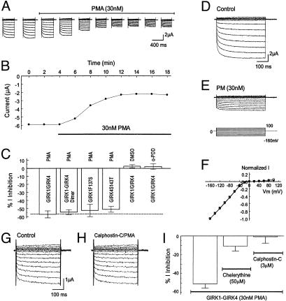
Inhibition of GIRK1/GIRK4 channels by PKC activation. (A) Whole-cell currents were studied in two-electrode voltage clamp by using a recording solution containing 90 mM K+. Inward rectifying currents were recorded from an oocyte 3 days after coinjection of the GIRK1 and GIRK4 cDNAs. Membrane potential (Vm) was held at 0 mV. A series of command pulse potentials from –160 mV to 100 mV with a 20-mV increment was applied to the cell. Exposure to 30 nM PMA produced the inhibition of GIRK currents. (B) The current amplitude in A measured at –160 mV was plotted against time. The currents decreased rapidly after the oocyte was exposed to 30 nM PMA. At maximal inhibition, the current amplitude was inhibited by ≈50%. (C) Summary of data obtained from PMA experiments. PMA (30 nM) inhibited heteromeric, dimeric, and homomeric GIRK channels to almost the same degree whereas DMSO and 4α-PDD had no effect on the heteromeric GIRK1/GIRK4. Data are shown as means ± SE (n = 4–9). (D–F) Voltage independence of GIRK1/GIRK4 inhibition by PMA. (D) Currents were recorded from an oocyte in the same condition as in A. These currents were strongly inhibited by exposure to 30 nM PMA (10 min). (F) When currents in D and E were scaled to the same magnitude at –160 mV, the I/V relationship of the currents recorded in these two conditions became identical. Open circles, control; filled triangles, PMA exposure. (G and H) Blockade of the PMA effect by specific PKC blocker calphostin-C (3 μM). (I) A similar effect was seen with another specific PKC blocker chelerythrine (n = 6–9).

