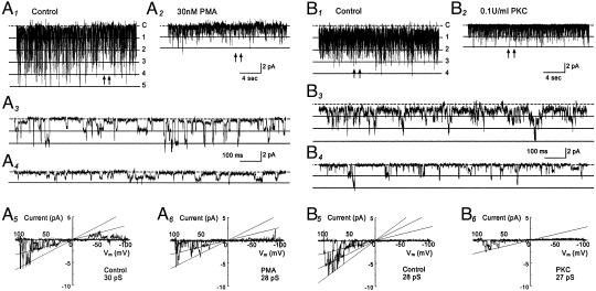
Fig. 2.
Comparison of the effect of PMA to that of PKC. (A1–A6) GIRK1-GIRK4 currents were recorded from a cell-attached patch with 150 mM K+ applied to the extracellular solution at a membrane potential of –80 mV. Five active currents were seen at baseline with NPo 0.671 (A1). These currents were inhibited when the oocytes was exposed to 30 nM PMA for 10 min (NPo 0.076, A2). (A3 and A4) The single-channel currents are better seen with an expended time scale taken from A1 and A2 at places pointed by arrows, respectively. (A5 and A6) In contrast to the NPo, single-channel conductance was inhibited only modestly by PMA. (B1–B6) Inhibition of the GIRK1-GIRK4 currents by purified catalytic subunit of PKC. (B1) Single-channel currents were studied in an excised inside-out patch with 150 mM K+ applied to both sides of the patch membrane. Four active channels are seen at baseline with NPo 1.143. (B2) The channel activity was strongly inhibited (NPo 0.144) 10 min after an exposure of the internal surface of the patch member to PKC (0.1 units/ml). (B5 and B6) PKC had very little effect on single-channel conductance in another inside-out patch. Note that substate conductances are seen in all records.

