Figure 1.
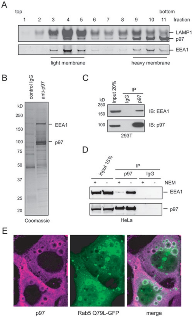
The p97 is localized to the endosomes and interacts with EEA1. (A) A post nuclear supernatant fraction from 293T cells was subject to sucrose gradient floatation to separate membranes based on their density. Collected fractions were analyzed by immunoblotting with the indicated antibodies. (B) A Coomassie blue stained gel showing proteins co-purified with p97 from solubilized cow liver membrane. (C) Co-IP analysis of EEA1-p97 interaction using 293T cell extracts. (D) As in C, except that HeLa cells were used. Where indicated, cell extracts were treated with 5 mM NEM. (E) Confocal analyses of COS7 cells showing endogenous p97 (left panel), overexpressed Rab5 (Q79L) GFP (middle) and the merged image (right).

