Figure 4.
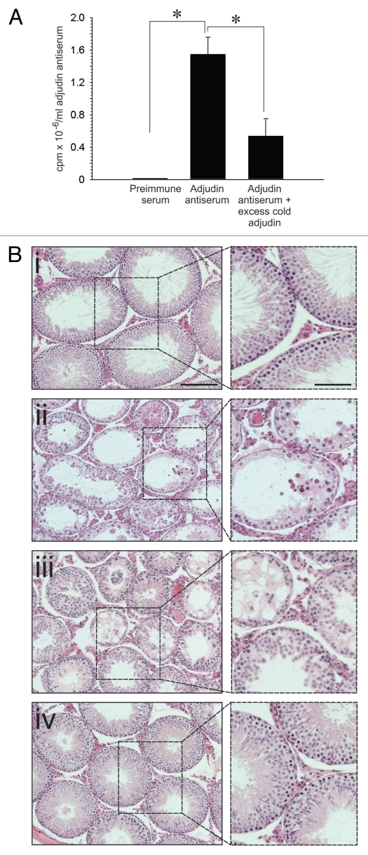
A study to assess the ability of the anti-adjudin antibody to protect adjudin-induced germ cell loss from the seminiferous epithelium in rat testes. (A) Adjudin competed with the binding of [3H]-adjudin to its antibody in a competitive binding assay. (B) Anti-adjudin IgG blocked the effects of adjudin on Sertoli-germ cell adhesion. (i) Cross-section of the control testis. (ii) Cross-section of the testis 21 days after treatment with adjudin (50 mg/kg b.w.). (iii) Cross-section of the testis 21 days after treatment with non-immune rabbit IgG and adjudin (50 mg/kg b.w.). (iv) Cross-section of the testis 21 days after treatment with anti-adjudin IgG and adjudin. All cross-sections were stained with H&E. Boxed areas in (i–iv) represent magnified views, and these are shown to the right of each image. Bar in (Bi) [also applies to (Bii–iv)] = 125 µm; bar in magnified views = 75 µm. Error bars represent mean ± SD from four different experiments. *p < 0.05. (two-way ANOVA followed by Tukey's post-hoc test).

