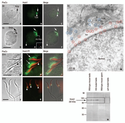
Figure 4.
Immunocytochemical localization and immunoblotting of Hook1. (A, D, G and J) are phase-contrast (PhaCo) microscopy. (A–C) (step 2 of spermiogenesis; S2): Hook1 immunoreactive sites are seen at the trans-Golgi network (TGN, white arrowhead in B) and the acrosome-acroplaxome of a rat round spermatid. The arrows in (A–C) indicate the Golgi. (C) is the merge of (A and B). (D–F) (S5): Hook1 is predominantly located at the acrosome-acroplaxome (Acr/Apx) region and in the centriolar region opposite to the acrosome (white arrowhead in E and F). The inset in (E) corresponds to a double staining with γ-tubulin (SIGMA, St. Louis, MO; working dilution: 1:50; catalog number T6557) to confirm the localization of Hook1 in the centrosome. The arrows in (D–F) indicate de Golgi. (F) is the merge of (D and E). (G–I) (S14): Hook1 is localized in the manchette (arrows in H) of elongating spermatids. No immunoreactivity is seen in the acrosome-acroplaxome region (arrowhead in G and H). PI denotes nuclear red staining with propidium iodide. (I) is the merge of (G and H). (J–L) (S19): Hook1 is seen in the HTCA (arrowheads in K) of mature spermatids. (L) is the merge of (J and K). (M) (S3): Immunogold electron microscopic localization of Hook1 in a rat round spermatid. The blue circles denote immunoreactive sites at the TGN region, mainly at the cytosolic side of proacrosomal vesicles. The red circles indicate the localization of Hook1 along the outer acrosomal membrane and at the inner acrosome membrane-acroplaxome (Apx) interface. Acr: acrosome. (N) Immunoblot showing the immunoreactive 92 kDa (729 amino acids) Hook1 protein in wild-type mouse testis, the acrosome-less Hrb mutant mouse testis, and wild-type mouse sperm but not in testis from the azh mutant, known to express truncated Hook1 lacking the C-terminus. Affinity purified polyclonal antibody was generated in rabbit using as antigen the peptide 625RNV IKT LDP KLN PAS639 corresponding to the C-terminal region of Hook1 (mouse; GeneBank Acc. No. AF487912). Hook1 antibody working dilution of 1:100 was used for indirect immunofluorescence and 1:25 for immunogold electron microscopy carried out according to previously reported procedures in reference 8. Scale bars in (A, D, G and J) 5 µm.

