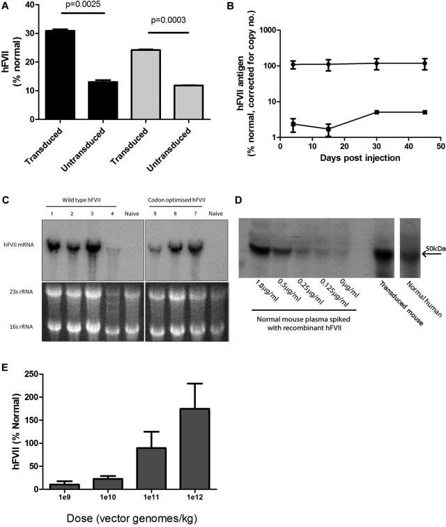
Figure 3.
Evaluation of AAV-mediated FVII expression. (A) FVII Ag (left side, ■) and activity (right side, ▩) in supernatant harvested from HepG2 cells 72 hours after the final transduction with scAAV8-LP1-hFVII-coop at an moi of 1 × 10⁵ vg/cell/d over 4 days. (B) Human FVII levels in plasma of male C57Bl/6 mice following a single tail vein administration of 2.8 × 10¹² vg/kg scAAV-encoding human FVII (circles = hFVII-coop, squares = hFVII-wt). (C) Top panels: Northern blot showing total human FVII mRNA levels in the liver of mice transduced with 2.8 × 10¹² vg/kg scAAV-LP1-FVII-wt (left) and scAAV-LP1-FVII-coop (right) compared with control untransduced animals (naive). Bottom image: Ethidium-stained agarose gel showing equal loading of liver RNA before transfer onto the nitrocellulose membrane. (D) Representative Western blot showing that human FVII-coop expressed following scAAV-mediated gene transfer (transduced mouse) has the same molecular weight as recombinant FVII and FVII detected in human pooled plasma. Serial dilution of recombinant FVII in naive mouse plasma suggests that the amount of human FVII in the experimental animal is > 1 μg/mL. (E) Human FVII levels in mice at 6 weeks after a single tail vein injection of between 1 × 10⁹-1 × 10¹² vg/kg scAAV-LP1-FVII-coop. Results are shown as means ± SEM.

