Figure 6.
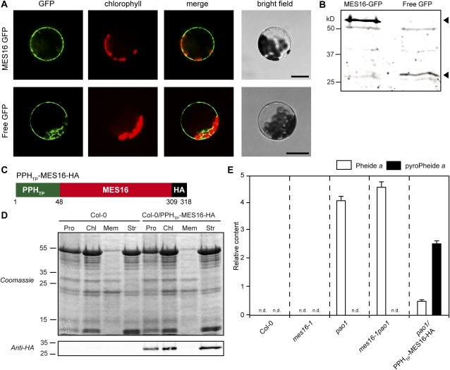
FCCs are the in vivo substrates of MES16. A, Transient expression of MES16-GFP and free GFP in Arabidopsis mesophyll protoplasts. GFP fluorescence (GFP) and Chl autofluorescence (chlorophyll) were examined by confocal laser scanning microscopy. The merge panels show overlays of GFP and autofluorescence. Bars = 20 μm. B, Anti-GFP immunoblotting of proteins from protoplasts expressing MES16-GFP and free GFP. The arrowheads indicate the predicted sizes of transiently expressed proteins. C, The chimeric construct used to target MES16 to the chloroplast. PPHTP, Amino acids 1 to 48 from PPH, representing the chloroplasts transit peptide; HA, HA tag. D, Verification of MES16 targeting to the chloroplast. Leaves of Col-0 and Col-0/PPHTP-MES16-HA were fractionated into protoplasts (Pro) and chloroplasts (Chl) and chloroplast subfractions (Mem, chloroplast membranes; Str, stroma). Gel loadings of protoplast and chloroplast fractions are based on equal amounts of chlorophyll. Anti-HA antibodies were used for detection of the chimeric protein. E, Quantification of Pheide a (white bars) and pyro-Pheide a (black bars) in dark-induced (5 d) Col-0, mes16-1, pao1, mes16-1pao1, and pao1/PPHTP-MES16-HA. Values are means of three replicates, and error bars represent sd. n.d., Not detected.

