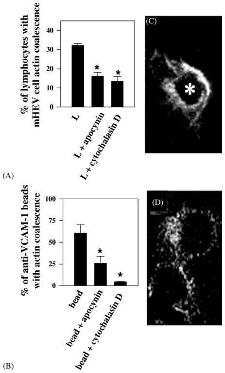
Fig. 6.
Lymphocytes and anti-VCAM-1-coated beads stimulate apocynin-inhibitable actin coalescence in endothelial cells at the site of contact. Lymphocytes and anti-VCAM-1-coated beads (2.5 × 10⁶/ml) were incubated in the presence or absence of 2.5 mM apocynin or 1 μM cytochalasin D with confluent monolayers of mHEVc cells for 5 min. The cells were fixed, permeabilized, labeled with TRITC-phalloidin, and examined by confocal microscopy. (A) Percentage of lymphocytes or (C) % of anti-VCAM-1-coated beads with endothelial cell actin coalescence at the site of contact with the lymphocytes or beads, respectively. L indicates lymphocytes. (*) P < 0.05 compared to controls (lymphocyte-stimulated or bead-stimulated mHEV cells). Micrographs shown are representative optical thin slices of TRITC-phalloidin labeled actin in the endothelial cell at the site of lymphocyte (C) or anti-VCAM-1-coated bead (D) contact. (*) in panel C, indicates the location of the center of a lymphocyte, above the thin slice shown, as determined by labeling with FITC-conjugated anti-CD45 (data not shown). There was no effect of bead or lymphocyte binding on the actin structure at the center of the mHEV cell (data not shown). Similar data were obtained for mHEVa cells. (Modified and reproduced with permission from Matheny et al., 2000. J. Immunol. 164:6550–6559.)

