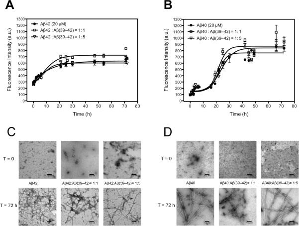
Figure 6.
The interference of fibril formation of Aβ42 and Aβ40 in the absence and presence of Aβ(39–42). A) Aβ42 (20 μM), 1:1 and 1:5 mixtures of Aβ42 and Aβ(39–42) and B) Aβ40 (20 μM), 1:1 and 1:5 mixtures of Aβ40 and Aβ(39–42) were incubated at 37 degree with agitation. β–sheet structure was monitored using ThT fluorescence C) and D) Electron microscopy photos of all samples mentioned above at time 0 and at 72 hours incubation. The scale bar represents 100 nm.

