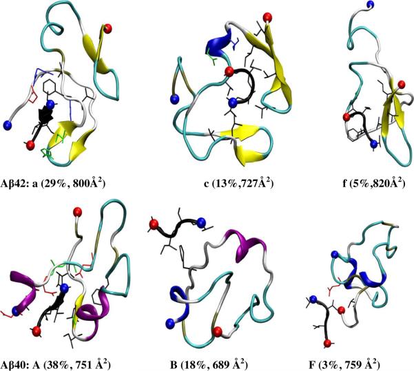
Figure 9.
Selected representative structures of Aβ·Aβ(39–42) complexes from the populated structural families (see Figure S9–S10 for structures from all families, a–f for Aβ42 and A–F for Aβ40). The abundance and collision cross section are noted below each structure. Only the side-chains in contact with Aβ(39–42) are shown (blue positively charged; red, negatively charged and black, hydrophobic). α-helical, 3–10-helical, β-extended, β-bridged, turn and coiled conformation are colored in purple, blue, yellow, tan, cyan and white, respectively. The positively charged N-termini and negatively charged C-termini are indicated by blue and red balls, respectively.

