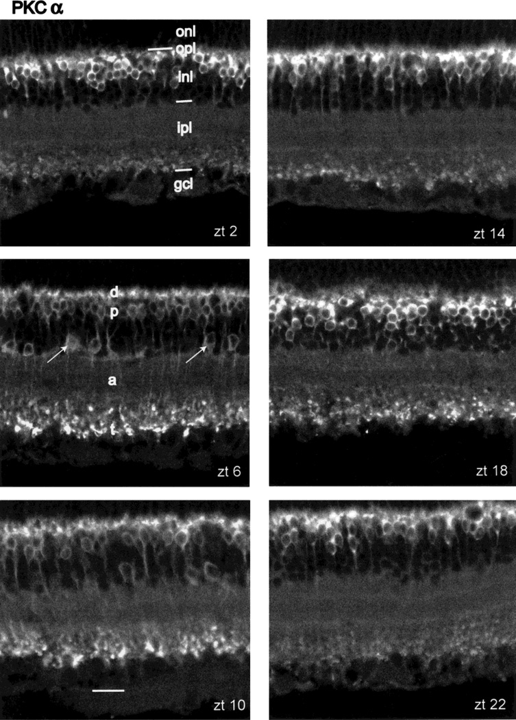
Fig. 1.
Diurnal variation in PKCα-IR. Panels illustrate vertical sections through central retina taken from rats sacrificed at ZT 02, 06, 10, 14, 18, and 22 hours, as labeled. All sections in this figure and the other figures are oriented with the photoreceptor layer up and the vitreous body below. The main neuronal class exhibiting PKC-IR is the rod bipolar cell. Its dendrites (d), cell bodies (p), axons (a), and axon terminals (t) are labeled in panel ZT 06. Amacrine cells showing PKC-IR are indicated by arrows in panel ZT 06. The layers of the retina are indicated in panel ZT 02 as follows: outer nuclear layer (onl), outer plexiform layer (opl), inner nuclear layer (inl), inner plexiform layer (ipl), and combined ganglion cell and optic fiber layer (gcl). Scale bar = 20 µm.

