Fig. 2.
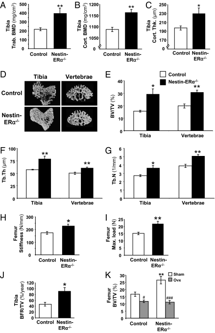
Deletion of ERα in nervous tissue results in increased bone mass. pQCT analysis of the tibia demonstrated increased trabecular volumetric bone mineral density (Tibia Trab. BMD) (A), increased cortical volumetric BMD (Tibia Cort. BMD) (B), and increased cortical thickness (Tibia Cort. Thk.) (C) in 3-mo-old nestin-ERα−/− mice compared with controls (n = 6). (D) Representative images of μCT analyses of trabecular bone in the proximal metaphysis of the tibia and in vertebrae L5. Bone volume per total volume (BV/TV) (E), trabecular thickness (Tb.Th) (F), and trabecular number (Tb.N) (G) of trabecular bone in the proximal metaphysis of the tibia and in vertebrae L5 (n = 5–8). Three-point bending of femur demonstrated increased (H) stiffness and (I) maximal load (Max. load) at failure in nestin-ERα−/− mice (n = 5). (J) Dynamic histomorphometric analysis of tibia giving bone formation rate per tissue volume (BFR/TV; n = 4–5). (K) Effect of ovx on bone mass in nestin-ERα−/− and control females. Three-month-old female mice were ovariectomized or sham-operated and terminated after 4 wk (n = 5–8). Values are given as mean ± SEM; *P < 0.05, **P < 0.01 vs. control; #P < 0.05, ###P < 0.001 vs. sham, Student's t test.

