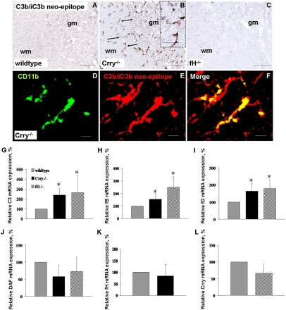
Fig. 2.
Alternative pathway activation in Crry−/− spinal cord. (A–C) Immunostaining for C3b/iC3b neoepitope, showing deposition in Crry−/− (B) but not in WT (A) or fH−/− (C) spinal cords. (D–F) Colocalization of C3b/iC3b with the microglial marker CD11b in Crry−/− mice. (Scale bar: A–C, 100 μm; D–F, 10 μm.) (G–L) Relative C3 (G), fB (H), fD (I), DAF (J), fH (K), and Crry (L) mRNA expression in spinal cords of WT (n = 7), Crry−/− (n = 7), and fH−/− (n = 4) mice. Values are normalized to the expression of β-actin and given as percentage (means ± SD) of WT control levels. Error bars are for n, number of animals. *P < 0.05 determined by one-way ANOVA.

