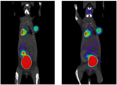
Fig. 4.
Small animal PET and CT images of a mouse bearing a lymphoma xenograft (right shoulder) after administration of [18F]FDG prepared on EWOD chip (left), and [18F]FDG prepared at the UCLA Biomedical Cyclotron facility (right). Injections and static scans (10 min duration, 1 h postinjection) were performed on the same mouse on consecutive days.

