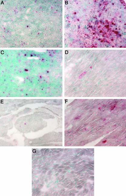
Fig. 1.
Histochemical evaluation of GUSB+ (red staining) cells. Liver (A), spleen (B), thymus (C), heart myocardium (D), and heart valve (E) from an 11.5-month-old treated MPSVII mouse. The myocardium from an adult +/+ mouse (F) and an untreated MPSVII mouse (G) are presented for comparison. (Magnification: ×120.)

