Fig. 3.
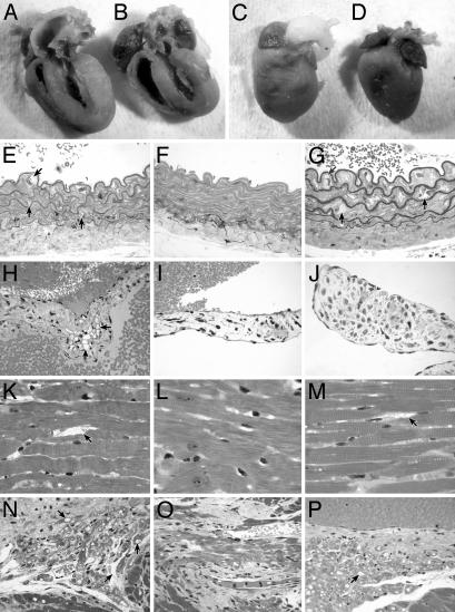
Gross and microscopic comparisons of heart pathology. (A–D) For gross pathology, tissues were fixed in formalin. The untreated MPSVII heart and left ventricle (A) are enlarged with respect to the +/+ heart (B). The untreated aortic root (C) is grossly distended when compared to the normal aortic root (D). The heart and ventricle in treated MPSVII mice are similar in size to the +/+ mice, but the aortic root distension is not corrected (data not shown). (E–P) Lysosomal storage is noted with an arrow. These sections compare lysosomal storage in various cardiac structures. (E) The wall of the untreated MPSVII aorta has irregular elastic lamina. There is extensive storage in the intimal, medial, and adventitial cells by comparison with the +/+ aorta (F). (G) The treated MPSVII aorta shows similar storage as in the untreated mouse aorta. The only reduction in storage occurred in adventitial cells. (H)An untreated MPSVII heart valve has abundant storage in stromal cells when compared to a normal heart valve (I) and a treated MPSVII valve (J). The heart valve stromal cells had less storage in the treated MPSVII mice. In the myocardium, untreated mice (K) have abundant storage in the perivascular and interstitial cells when compared to those in +/+ (L) and treated (M) mice. The interstitial cells and myocytes of the conduction system in untreated mice (N) have storage when compared to those in normal mice (O). The interstitial cells but not the conduction tissue myocytes are cleared of storage in the treated MPSVII mice (P). (Magnification: ×100.)

