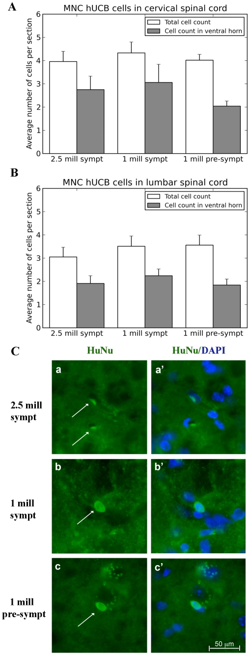
Figure 3. Distribution of MNC hUCB cells in the spinal cord of G93A mice.
Administered MNC hUCB cells were identified immunohistochemically by a human-specific marker (HuNu) in the spinal cord of cell-treated mice at 17 weeks of age, 4 weeks (symptomatic) or 8 weeks (pre-symptomatic) post-transplant. In the total area of cervical (A) and lumbar (B) cervical spinal cord, HuNu positive MNC hUCB cells were found irrespective (p>0.05) of injected cell doses or time beginning treatment. In all cell-treated mice, more than 50% of the observed cells were in ventral horn gray matter. (C) Immunohistochemical staining of MNC hUCB cells in the lumbar spinal cord. MNC hUCB cells positive for HuNu (green, arrow) were detected in the lumbar spinal cord of mice receiving 2.5×10⁶ (a) or 1×10⁶ (b) cells symptomatically or 1×10⁶ cells pre-symptomatically (c). Cells were frequently observed inside the capillary lumen, but also in the spinal cord parenchyma. (a′), (b′), and (c′) are merged images with DAPI. Scale bar: a–c′ is 50 µm.

