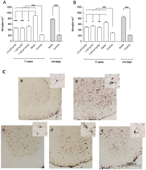
Figure 6. Characteristics of microglial cells in the spinal cords of G93A mice.
Microglial densities were measured in the cervical (A) and lumbar (B) ventral horns of G93A mice at 17 weeks of age and at end-stage of disease. Microglial densities were significantly (p<0.001) higher in Media-injected mice (Gr 4) compared to control mice (Gr 5) of the same ages. MNC hUCB cell administrations significantly (p<0.001) decreased the number of microglia in the spinal cord of G93A mice compared to Media mice. No significant differences were detected between the cell-treated groups. *p<0.05, **p<0.01, ***p<0.001. (C) Immunohistochemical staining of microglia in the lumbar spinal cord at 17 weeks of age. Microglial cells stained for anti-Iba-1 antibody were sparse in controls (a) and microgliosis was noted in Media-treated animals (b). MNC hUCB cells decreased microglial density in mice from Gr 1 (c), Gr 2 (d), and Gr 3 (e). Morphological analysis of microglial cells determined numerous activated cells with large cell bodies and short processes in Media-injected mice, whereas ramified microglia were mostly observed in cell-treated animals, particularly in Gr 1 and Gr 3 mice and controls (inserts in a–e). Scale bar: a–e is 200 µm; in a–e inserts is 25 µm.

