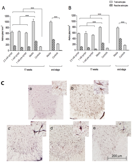
Figure 7. Characteristics of astrocytes in the spinal cord of G93A mice.
Astrocyte densities were measured in the cervical (A) and lumbar (B) ventral horns of G93A mice at 17 weeks of age and at end-stage of disease. Astrocytic densities were significantly (p<0.001) higher in Media-injected mice (Gr 4) at 17 weeks of age and at end-stage of disease vs. controls (Gr 5) of the same ages. Significant (p<0.001) decrease in the number of astrocytes was determined in cell-treated G93A mice compared to Media mice. No significant statistical differences were detected between the cell-treated groups. Higher number of reactive astrocytes was observed in Media-injected mice at 17 weeks of age and at end-stage disease compared to cell-treated animals. ***p<0.001. (C) Immunohistochemical staining of astrocytes in the lumbar spinal cord of G93A mice at 17 weeks of age. Anti-GFAP antibody staining showed low astrocyte density in controls (a) and astrocytosis in Media-treated animals (b). Considerably decreased actrocytic density was observed in mice from Gr 1 (c), Gr 2 (d), and Gr 3 (e). Astrocyte cell reactivity was also reduced in cell-treated mice vs. Media-injected mice (inserts in a–e). Scale bar: a–e is 200 µm; in a–e inserts is 25 µm.

