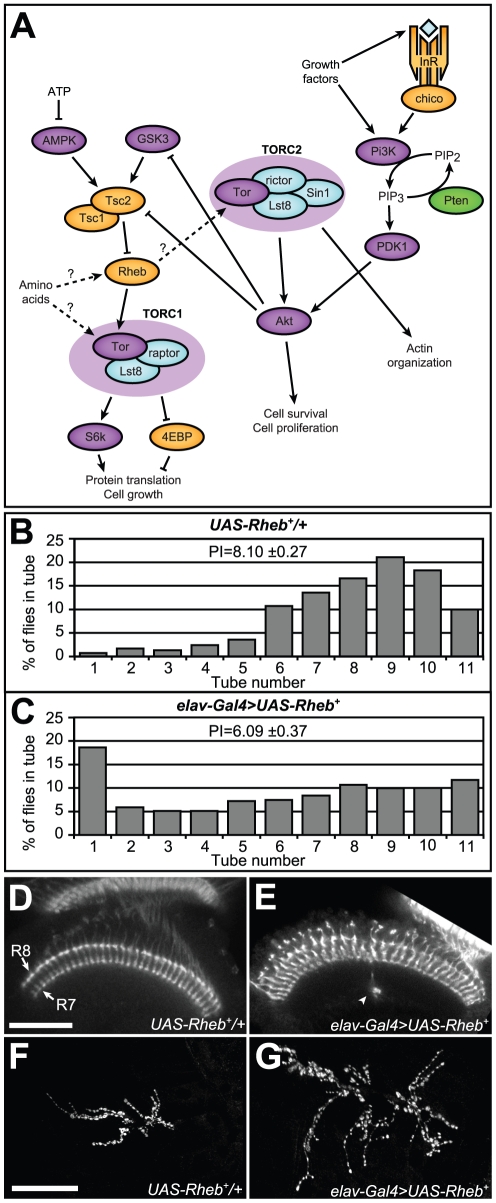
Figure 1. Rheb overexpresion causes defects in behavior, axon guidance, and synapse morphology.
(A) Schematic diagram of the Tor pathway. Kinases are purple, Tor-complex components are blue, phosphatases are green, and other components are orange. The Tsc1/2 heterodimer inhibits Rheb, which in turn controls the activity of two Tor-containing complexes, TORC1 and TORC2. Relationships which are not fully-understood or could have multiple intermediary steps are shown as dashed arrows with question marks. (B, C) Phototaxis measurements in flies overexpressing Rheb in all neurons (elav-Gal4>UAS-Rheb+, n = 416) compared to control flies that have UAS-Rheb + transgenes but lacked the neuron-specific Gal4 driver (UAS-Rheb+/+, n = 531). The percentage of flies in each tube of the phototaxis apparatus after 10 trials is shown, along with the phototaxis index (PI), a cumulative score where higher numbers indicate a stronger phototaxis response. (D, E) Optic lobes of pupal brains oriented with retinas toward the top of the photos. Visualization of photoreceptor projections with Anti-Chaoptin antibody staining reveals that R7 and R8 photoreceptor neurons form two highly-structured parallel rows of terminations in the medulla (arrows), with no axon bundles projecting beyond the R7 termination layer in controls. (E) Rheb overexpression in neurons caused some axon bundles to continue growing beyond their proper R7/R8 termination targets to eventually stop elsewhere within the medulla (arrowheads). (F, G) Anti-CSP (Cysteine string protein) staining at the larval NMJ reveals synaptic active zones known as boutons. Neuron-specific overexpression of Rheb resulted in neuromuscular synapses approximately 50% larger than corresponding synapses in animals lacking a Gal4 driver. Scale bars are 50 microns.

