Vascular endothelial growth factor (VEGF) is upregulated in multiple myeloma (MM), and circulating VEGF levels may correlate with response to therapy (Hideshima, et al 2005, Pittini, et al 2002). Thalidomide has been part of the standard treatment for MM and is thought to inhibit VEGF-associated angiogenesis (Du, et al 2004). Bevacizumab, a monoclonal antibody directed against VEGF-A, inhibits VEGF (Jenab-Wolcott and Giantonio 2009). Accordingly, we set out to test the efficacy and safety of bevacizumab alone and in combination with thalidomide in MM patients.
A phase II prospective randomized/stratified trial led by the California Cancer Consortium, and including the University of Chicago, was approved by the Cancer Therapy Evaluation Program/ National Cancer Institute of the National Institutes of Health (N01-CM-62209). Patients with prior thalidomide exposure received bevacizumab alone (Arm A). Thalidomide-naïve patients were randomized to either arm B (bevacizumab alone) or C (combination therapy). The study was closed early due to poor accrual, attributable to competing trials providing access to lenalidomide and bortezomib (Knight 2005, Lu, et al 2009, Moschetta, et al 2010).
The primary objectives were response rate, event-free survival, and toxicity. The secondary objective was to measure markers of angiogenesis and assess any correlation with outcome. Immunohistochemical (IHC) staining of VEGF (VG-1, Neomarkers, Freemont, CA) and its two receptors, VEGFR1/Flt-1 (AB-1, Neomarkers) and VEGFR2/KDR (AB-1, Neomarkers) was carried out on bone marrow clots or cores obtained at baseline.
The study was conducted between October 2001 and November 2004. Patients aged 18 years or older, with relapsed/progressive MM and a Karnofsky performance status (KPS) > 60%were enrolled. All patients signed a voluntary informed consent form, approved by the institutional review boards of the participating institutions.
Bevacizumab was given at 10 mg/kg intravenously over a 90-min period every 14 days. Thalidomide was escalated from 100 mg/day by 100 mg/week, up to 400 mg/day. Treatment cycle length was 56 days. Treatment was discontinued due to disease progression, development of grade 3 or 4 toxicities that did not resolve to grade 1 or less (maximum 3 weeks’ delay was allowed), non-compliance, or patient request or physician discretion. The National Cancer Institute’s Common Toxicity Criteria version 2.0 (http://ctep.cancer.gov/protocolDevelopment/electronic_applications/docs/ctcv20_4-30-992.pdf) was used for toxicity and adverse event reporting.
Complete response was defined as disappearance of the paraprotein in the serum and/or urine by immunofixation and less than 5% plasma cells on bone marrow evaluation. A partial remission was defined as a ≥ 50% reduction but still detectable level of paraprotein, and if present, a ≥ 50% reduction in urine M-component. Stable disease was defined as < 50% reduction in paraprotein, or if the patient had light-chain disease only, a > 50% reduction in the urine M-component (Bence-Jones protein). Progressive disease was defined as a 25% increase in paraprotein from the lowest level observed, measured on at least 2 separate occasions two weeks apart. We defined event-free survival (EFS) as synonymous with time to treatment failure (TTF) to avoid reporting artificially long progression-free survival in patients who declined further protocol therapy prior to progression. TTF was therefore defined as the time from the first day of treatment to the first observation of disease progression, death, or treatment cessation due to toxicity or patient refusal.
Fourteen patients consented; one withdrew prior to initiation of treatment, and another became ineligible due to a drop in KPS. Twelve evaluable patients, 8 female, 4 male, (median age: 58 years, range: 50–75) were enrolled; six received bevacizumab alone (Arms A or B); six received combination therapy (Arm C). Eight of the patients were enrolled with stage III disease (Durie and Salmon 1975) and two each with stages I and II. The median β2 microglobulin was 2.7 mg/l (range 1.0–9.9 mg/l) with 9 cases of IgGК, 2 patients with IgGλ, and 1 case of non-secretory myeloma. Previous treatments included VAD (vincristine, doxorubicin, dexamethasone), thalidomide, melphalan, and prednisone, received by10, 3, 5, and 3 patients respectively, with 10 patients receiving other agents. No patient received bortezomib or lenalidomide. The median number of prior systemic regimens was 3 (range 0–5). Five patients had undergone radiation therapy; 7 had undergone autologous transplantation.
Toxicities were mild: The combination therapy resulted in grade 3 lymphopenia (n=1), fatigue (n=1), and grade 4 pulmonary hypertension (n=1; early cessation due to shortness of breath in a patient with prior exposure to phen-phen). Bevacizumab-associated grade 3 toxicities were fatigue (n=1), hypertension (n=1), neutropenia (n=1), and hyponatraemia (n=1).
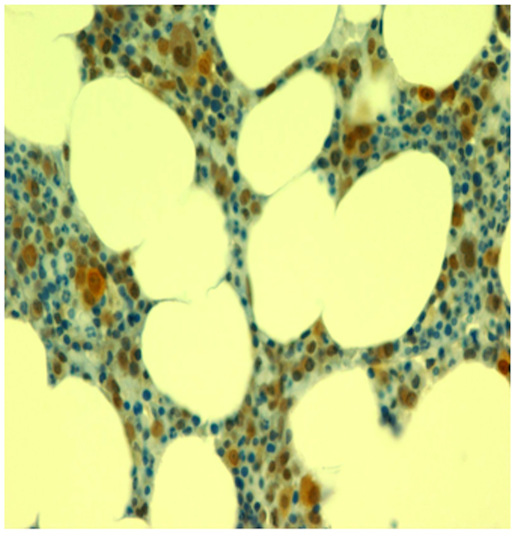
Plasma cell expression of VEGF was observed in 7 of 9 MM patients examined by IHC, appearing as a diffuse, cytoplasmic pattern varying in intensity from moderate (++) to strong (++++). VEGF staining in the lymphoblastic or erythroblastic lineages was not observed whereas occasional staining of polymorphonuclear cells was seen. Five of 9 samples displayed VEGFR1 and 4 expressed VEGFR2, but receptor staining was weak (+) to moderate and restricted to myeloid and monocytic cells. When VEGFR1 or 2 receptors were observed by IHC, there was a 100% concordance with VEGF expression.
Patient responses and outcomes are summarized in Table I. On Arm C (combination therapy), 2 patients achieved a partial response for 224 days and 369 days, and 3 experienced stable disease (223, 228, and 350 days), with a median EFS of 287 days (range: 37–369). Among those who received bevacizumab alone, one patient – with the greatest expression of VEGF (Figure 1) on MM cells – achieved stable disease for 238 days, but the median EFS for the cohort was only 49 days (range: 29–238).
Table I.
Event-free survival by treatment arm
| Patient | Best response | TTF (days) |
|---|---|---|
| Arm A: Thalidomide-exposed patients treated with bevacizumab | ||
| 1 | PD | 42 |
| 2 | PD | 29 |
| 3 | PD | 56 |
| Arm B: Thalidomide-naïve patients treated with bevacizumab | ||
| 1 | PD | 69 |
| 2 | PD | 36 |
| 3 | SD | 238 |
| Arm C: Thalidomide-naïve patients treated with the combination | ||
| 1 | SD | 37* |
| 2 | SD | 223 |
| 3 | SD | 228 |
| 4 | PR | 224** |
| 5 | PR | 369 |
| 6 | SD | 350 |
Early cessation of therapy due to pulmonary hypertension-related shortness of breath in a patient with prior exposure to diet pills (phen-phen), whose symptoms resolved and was able then to take thalidomide;
Patient came off therapy and went on to receive an autologous transplant
PD: progressive disease; SD: stable disease; PR: partial response
Figure 1.
VEGF expression on myeloma cells in the bone marrow of a patient treated with bevacizumab and thalidomide (1000× amplification)
The observed VEGF or VEGF receptor expression patterns support a paracrine role of VEGF in MM as demonstrated in preclinical models. Low accrual prevented correlation of VEGF or VEGFR1/VEGFR2 expression with response. Combination therapy, in this limited sample, yielded similar results to single agent thalidomide. Future clinical trials exploring the role of VEGF or VEGFR inhibition need to focus on patients whose myeloma cells are enriched for VEGF expression, and such trials are likely to be successful only if they involve combinations including agents with proven efficacy (Ribatti and Vacca 2003, Roccaro, et al 2006).
Acknowledgements
Supported by NCI Grants 33572 and Contract N01-CM-62209
Footnotes
Presented at the 47th Annual Meeting of the American Society of Hematology (ASH), Atlanta, GA, December 10–13, 2005.
REFERENCES
- Du W, Hattori Y, Hashiguchi A, Kondoh K, Hozumi N, Ikeda Y, Sakamoto M, Hata J, Yamada T. Tumor angiogenesis in the bone marrow of multiple myeloma patients and its alteration by thalidomide treatment. Pathol Int. 2004;54:285–294. doi: 10.1111/j.1440-1827.2004.01622.x. [DOI] [PubMed] [Google Scholar]
- Durie BG, Salmon SE. A clinical staging system for multiple myeloma. Correlation of measured myeloma cell mass with presenting clinical features, response to treatment, and survival. Cancer. 1975;36:842–854. doi: 10.1002/1097-0142(197509)36:3<842::aid-cncr2820360303>3.0.co;2-u. [DOI] [PubMed] [Google Scholar]
- Hideshima T, Podar K, Chauhan D, Anderson KC. Cytokines and signal transduction. Best Pract Res Clin Haematol. 2005;18:509–524. doi: 10.1016/j.beha.2005.01.003. [DOI] [PubMed] [Google Scholar]
- Jenab-Wolcott J, Giantonio BJ. Bevacizumab: current indications and future development for management of solid tumors. Expert Opin Biol Ther. 2009;9:507–517. doi: 10.1517/14712590902817817. [DOI] [PubMed] [Google Scholar]
- Knight R. IMiDs: a novel class of immunomodulators. Semin Oncol. 2005;32:S24–S30. doi: 10.1053/j.seminoncol.2005.06.018. [DOI] [PubMed] [Google Scholar]
- Lu L, Payvandi F, Wu L, Zhang LH, Hariri RJ, Man HW, Chen RS, Muller GW, Hughes CC, Stirling DI, Schafer PH, Bartlett JB. The anti-cancer drug lenalidomide inhibits angiogenesis and metastasis via multiple inhibitory effects on endothelial cell function in normoxic and hypoxic conditions. Microvasc Res. 2009;77:78–86. doi: 10.1016/j.mvr.2008.08.003. [DOI] [PubMed] [Google Scholar]
- Moschetta M, Di Pietro G, Ria R, Gnoni A, Mangialardi G, Guarini A, Ditonno P, Musto P, D'Auria F, Ricciardi MR, Dammacco F, Ribatti D, Vacca A. Bortezomib and zoledronic acid on angiogenic and vasculogenic activities of bone marrow macrophages in patients with multiple myeloma. Eur J Cancer. 2010;46:420–429. doi: 10.1016/j.ejca.2009.10.019. [DOI] [PubMed] [Google Scholar]
- Pittini V, Teti D, Arrigo C, Aloi G, Righi M. Thalidomide treatment of relapsed multiple myeloma patients and changes in circulating VEGF and bFGF. Br J Haematol. 2002;119:275. doi: 10.1046/j.1365-2141.2002.37694.x. [DOI] [PubMed] [Google Scholar]
- Ribatti D, Vacca A. On the use of thalidomide as an antiangiogenic agent in the treatment of multiple myeloma. Ann Hematol. 2003;82:262. doi: 10.1007/s00277-003-0622-4. [DOI] [PubMed] [Google Scholar]
- Roccaro AM, Hideshima T, Raje N, Kumar S, Ishitsuka K, Yasui H, Shiraishi N, Ribatti D, Nico B, Vacca A, Dammacco F, Richardson PG, Anderson KC. Bortezomib mediates antiangiogenesis in multiple myeloma via direct and indirect effects on endothelial cells. Cancer Res. 2006;66:184–191. doi: 10.1158/0008-5472.CAN-05-1195. [DOI] [PubMed] [Google Scholar]


