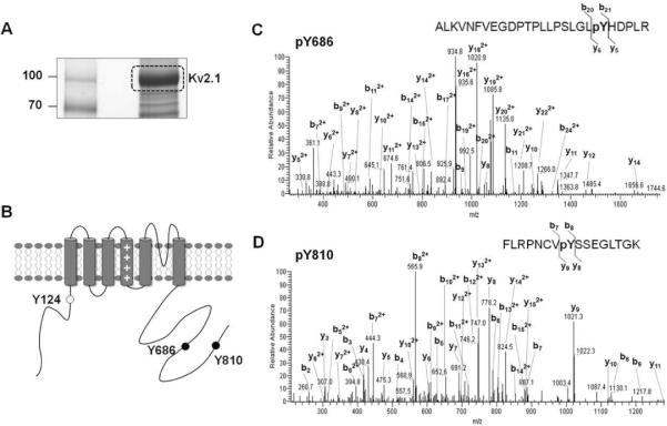
Fig. 2.
Identification of Kv2.1 tyrosine phosphorylation sites Y686 and Y810. (A) Immunopurification of Kv2.1. Coomassie brilliant blue-stained SDS gel of a large-scale immunopurification showing the yield of Kv2.1 channel. (B) Cartoon of the membrane topology of Kv2.1 showing tyrosine phosphorylation sites identified by LC-MS/MS analyses. Black dots mark the novel tyrosine phosphorylation sites identified in this study; white dots mark tyrosine phosphorylation sites identified in a previous study. (C) MS/MS spectrum of Kv2.1 tyrosine phosphopeptide pY686. Shown is the product ion spectrum of a triply charged, singly phosphorylated tryptic peptide at m/z 982.20. (D) MS/MS spectrum of Kv2.1 tyrosine phosphopeptide pY810. Shown is the product ion spectrum of a triply charged, singly phosphorylated tryptic peptide at m/z 637.34.

