
Figure 1.
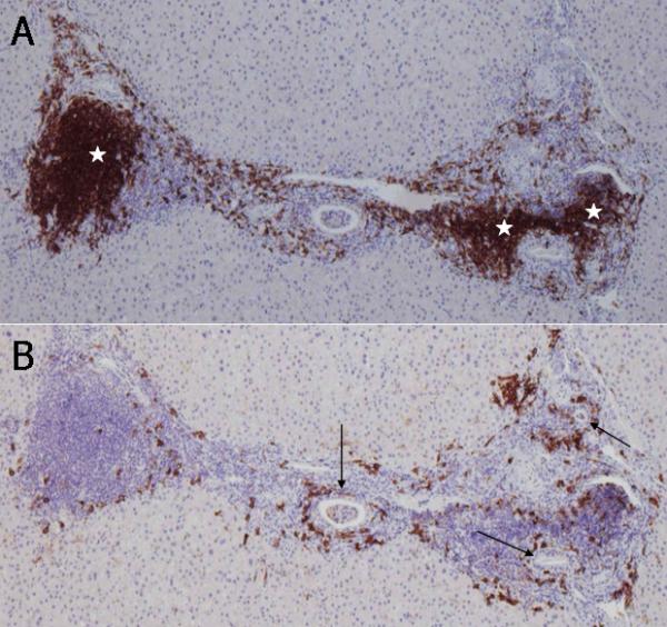
Immunohistochemical staining of CD20+ B lymphocytes and CD38+ cells in consecutive sections of a PBC liver. A. CD20+ B lymphocytes are either aggregated in lymph follicle-like structures (white stars) or scattered around inflamed portal tracts. B. Coronal arrangement (CA) of CD38+ cells surrounding the intralobular bile ducts (arrows). Note that CD38+ cells are scarce in lymph follicle-like structures. ABC method, x10.