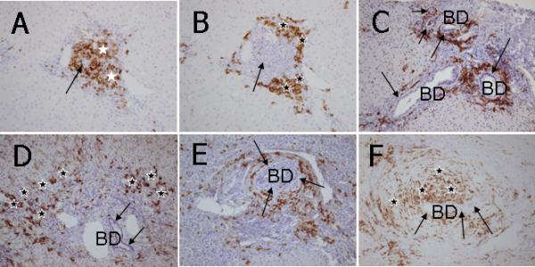
Figure 3.

Immunohistochemical staining of CD20+ and CD38+ cells in liver sections. A. In CH-C, CD20+ B lymphocytes are aggregated in a follicle-like structure in an inflamed portal tract (white stars). Note that an intrahepatic bile duct (an arrow) is centered in such a follicle-like structure. B. In a consecutive section of CH-C liver (with panel A), CD38+ cells are primarily located in the periphery of inflamed portal tracts (black stars) apart from the intrahepatic bile ducts (an arrow). C. In AIH, CD38+ cells are not observed in the proximity of intrahepatic bile ducts (BD, arrows) but abundantly infiltrated in the area of interface hepatitis (black stars). D. In PBC, CD38+ cells show the coronal arrangement surrounding intrahepatic bile ducts (BD) with CNSDC (arrows). E. In a case of stage 4 PSC, CD38+ cells are surrounding the onion skin-like fibrosis that is a characteristic of PSC (arrows). This pattern is coined a satellite-like arrangement (SA) of CD38+ cells surrounding concentric periductal fibrosis. The SA pattern is different from the CA of CD38+ cells in PBC located just beneath the cholangioepithelium with CNSDC, as shown in Figures 2C and 4D. F. In a case of stage 1 PSC, CD38+ cells are concentrically scattered in the onion skin-like fibrosis (black stars) surrounding an intrahepatic bile duct (arrows). This pattern is also regarded as SA of CD38+ cells. ABC method, x25.
