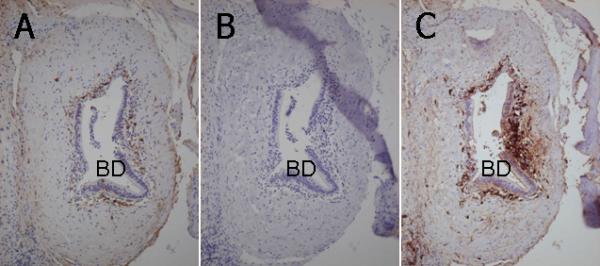
Figure 4.

Immunohistochemical staining of IgG, IgA and IgM in consecutive liver sections of a PBC liver. A. IgG+ cells demonstrate a relatively loose coronal arrangement surrounding an intrahepatic bile duct (BD) with CNSDC. B. IgA+ cells are not found in the vicinity of this intrahepatic bile duct (BD) with CNSDC. C. IgM+ cells demonstrate a dense and distinct coronal arrangement surrounding this intrahepatic bile duct (BD) with CNSDC. Labeled streptavidin-biotin method, x25.
