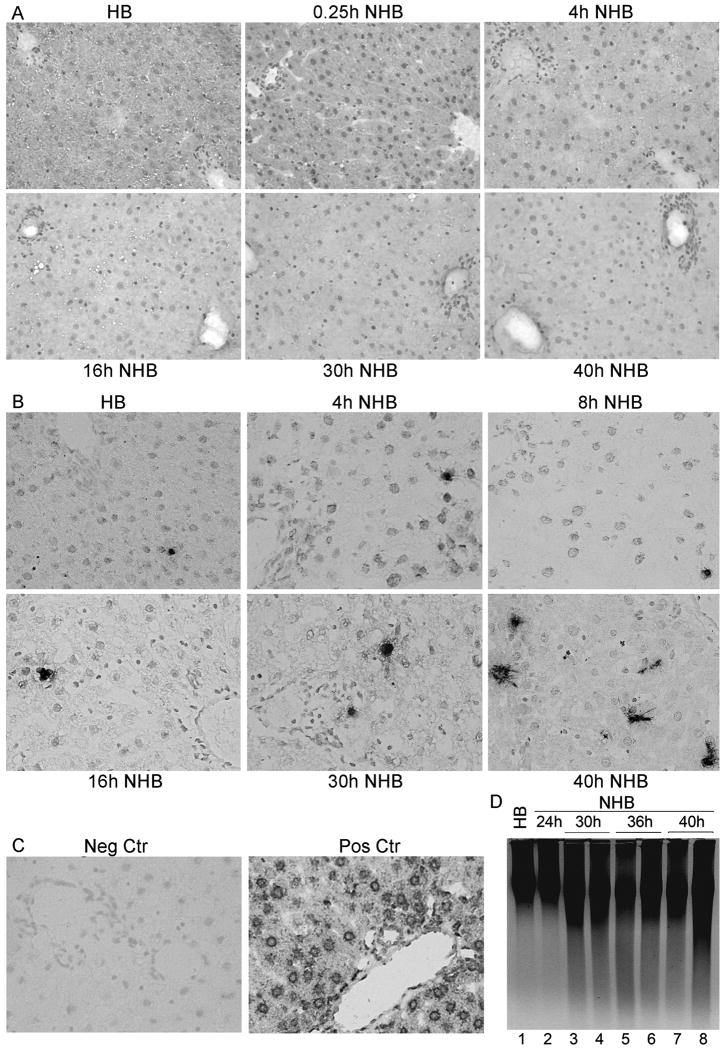
Figure 1. Integrity of liver in NHB donors.
(A) Hematoxylin and eosin-stained sections from HB and NHB donors after death, as indicated, showing tissues were intact without necrosis or autolysis. (B) TUNEL+ cells were infrequent in HB and NHB donor livers. (C) Negative control liver without TUNEL and DNAse-treated control liver with extensive TUNEL. (D) DNA laddering showing little hepatic apoptosis over time. Orig. Mag., A and B, ×400; B, methylgreen counterstain.

