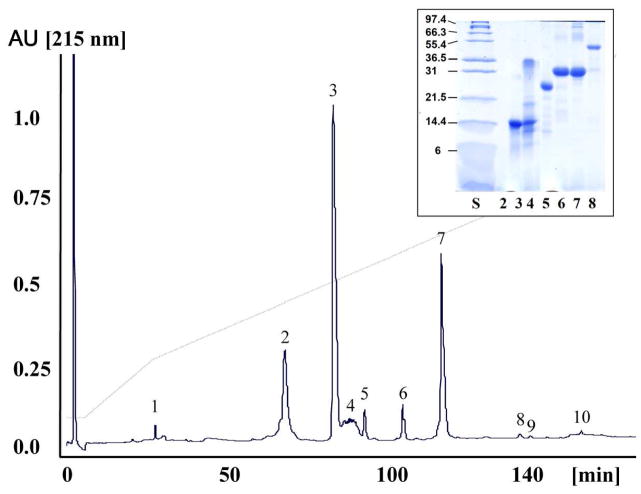
Fig. 1. Characterization of the venom proteome of Crotalus tigris.
Reverse-phase HPLC separation of the venom proteins of C. tigris. Insert, SDS-PAGE of the reverse-phase HPLC separated venom proteins run under reduced conditions. Molecular mass markers (in kDa) are indicated at the side of each gel. Protein bands were excised and characterized by mass fingerprinting and CID-MS/MS of selected doubly- or triply-charged peptide ions (Table 1).

