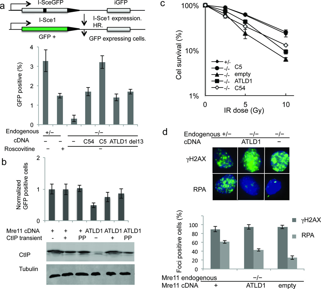
Figure 5. CtIP DSB repair functions are dependent on the Mre11 C-terminus.
(a) Schematic of DR-GFP assay for DSB repair by HR (top). Comparison of HR repair in MEFS of the indicated Mre11 genotypes (bottom). Error bars are average ± s.d. of at least 3 experiments.
(b) Complementation of HR deficiency by transient expression of CtIP (top). All MEFs were Mre11−/− at the endogenous locus. cDNAs expressed are indicated below the bar graph. Stable Mre11 expression was either wild–type (+) or ATLD1. Transient CtIP expression was either wild–type CtIP (+) or phosphomimetic mutant CtIP (pp). Western blot indicates levels of total CtIP protein (bottom). Tubulin is loading control.
(c) IR sensitivities of MEFS with Mre11 endogenous (left) and cDNA (right) genotypes indicated in the panel. Error bars are average ± s.d. of at least 3 experiments.
(d) Immunoflourescent foci induced by IR (10 Gy) (above). γH2AX (top) marks DSBs and RPA (bottom) indicates resection. Quantitation of foci is at bottom. Error bars are average ± s.d. of 3 experiments. Foci-positive is defined as cells with >10 foci.

