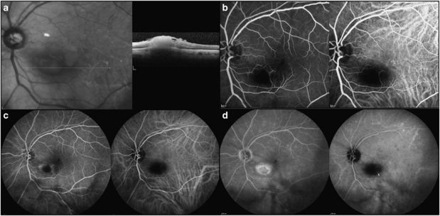
Figure 2.
Imaging studies of toxoplasmic focal chorioretinitis. (a) OCT image through lesion showing inner retinal hyperreflectivity with shadowing of the outer retina and choroid. (b) Early angiogram, 50 s. There is hypofluorescence of the lesion in both the fluorescein (left) and the indocyanine angiogram (right). (c) Mid-phase of the angiogram, 2.33 min. Hyperfluorescence begins at the edge of the focal lesion. The ICG remains hypofluorescent. (d) Late angiogram, 15.11 min. The lesion is almost fully stained with fluorescein. In the ICG the lesion is hypofluorescent and surrounded by dark dots most visible in the temporal macula.

