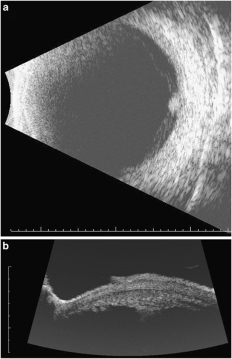
Figure 4.
Persistent fungal endophthalmitis following pars plana vitrectomy and removal of infected capsular bag. During a second surgery the entire bag and lens implant were removed, but inflammation persisted. Ultrasound confirmed focal deposits in the ciliary body region in the meridian where the infected capsular plaques had been noted. (a) Anterior to the equator at 3:00 (3EA) there is a focal deposit in the ciliary body region. (b) Anterior segment B scan confirms a ciliary body deposit at the 3:00 position (3T). Notice the ciliary processes to the left of the deposit. At surgery, a focal white deposit was found between 3 and 4:00 adherent to the ciliary processes. Removal of it with the vitreous cutter and picks followed by repeated injections of amphotericin enabled the infection to be cured.

