Figure 6.
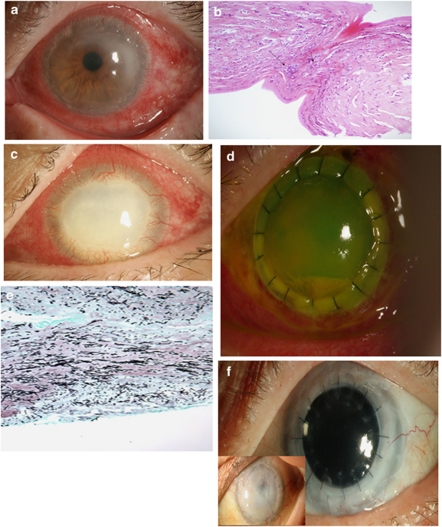
Photos represent the patient presented as Case no. 3. Photo (a) shows the clinical presentation as a ‘ring infiltrate' type of keratitis. This photo demonstrated how the patient presented to our institution. Photo (b) demonstrated the results of the biopsy that are compatible with Acanthamoeba keratitis. The patient lost to follow-up and a month later presented with significant corneal melt and worsening keratitis (Photo c). The patient was taken to the operating room for a therapeutic corneal transplant and photo (d) shows the patient's cornea one day after graft was performed. Photo (e) demonstrated the histology results done for the cornea after the graft compatible with fungal keratitis. Photo (f) shows the patients cornea after graft at later time points. Anterior photo demonstrates graft failure 2 months after the primary intervention and the posterior photo shows a clear cornea 8 months after optical penetrating keratoplasty.

