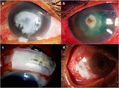
Figure 1.
Fungal infections of eye. (a) Shows fungal keratitis with dry gray to dirty white infiltrate and raised edges. (b) Shows keratitis with pigmented infiltrate. (c) Shows pigmented fungal growth on the surface of scleral buckle in a case of fungal scleritis. (d) Shows ulcerated scleritis lesion.

