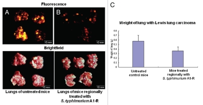
Figure 5.
S. typhimurium A1-R inhibition of Lewis lung carcinoma experimental metastasis with intrathoracic bacteria administration. Female C57 immunocompetent mice, aged 6 weeks, were injected with RFP-expressing Lewis lung carcinoma cells (1 × 10⁶ in 100 µl PBS) into the tail vein. Beginning at day 5, 1 × 10⁸ CFU S. typhimurium were injected into thoracic cavity weekly for three weeks. All animals were sacrificed at week 4. The excised lungs were weighed and imaged using the Olympus OV100. (A) RFP-labeled metastasis in the lungs of untreated C57 control mice. (B) RFP-labeled metastasis in the lungs of mice treated by intrathoracic S. typhimurium A1-R-GFP. (C) The weight of the lungs from mice treated with intrathoracic S. typhimurium A1-R compared with the weight of the lungs from untreated mice.

