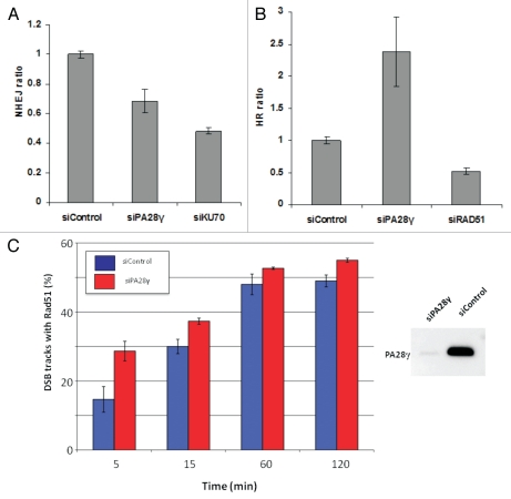
Figure 4.
Effect of PA28γ depletion on the NHEJ and HRR pathways of DSB repair. The experimental systems are based on cells in which interrupted GFP-encoding sequences containing recognition sites of the rare cutter restriction endonuclease I-SceI were incorporated into the cellular genome. In one system, the repair of I-SceI-induced DSB via HRR regenerates an active GFP-encoding sequence,69,71 and in the other, this effect is obtained following NHEJ-mediated repair of the break.70 In both cases, the GFP signal is monitored using FACS. (A) Effect of PA28γ depletion on NHEJ. HeLa cells containing the reporter sequences for NHEJ39,70 were transfected with an I-SceI-encoding plasmid along with the indicated siRNA oligonucleotides and analyzed 72 h later by flow cytometry. GFP-positive cells are gated, and the percentage of GFP-positive cells in PA28γ-depleted cells is normalized against that of cells transfected with irrelevant siRNA (siLuciferase). Cells depleted of the KU70 protein, a major NHEJ player, served as a positive control. Shown is the mean of the NHEJ ratio (average of triplicates). Error bars represent standard error. Results of one of three independent experiments are shown. (B) Similar analysis in U2OS cells containing the HRR reporter.69 Cells depleted of the RAD51 protein, a major HRR player, served as positive control. Results of one of four independent experiments are shown. Error bars represent standard deviation. (C) Quantification of the accumulation of RAD51 at DSBs in the presence or absence of PA28γ. U2OS cells were transfected with siRNAs against luciferase (siControl) or PA28γ, cultured for 48 h and irradiated with α-particles as described in references 40 and 68 to produce linear tracks of DSBs. The effectiveness of the downregulation was analyzed by immunoblotting, as displayed on the right. At the indicated time points after irradiation, the cells were stained for DNA (DAPI), γH2AX and RAD51. The γH2AX staining marked the tracks of DSBs, and the percentage of DSB tracks that co-localized with RAD51 was determined for 100 DSB track positive cells per data point. Error bars represent the SEM of three independent experiments.

