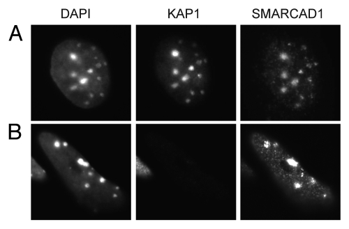
Figure 6.
Localization of SMARCAD1 to pericentric heterochromatin is not dependent on KAP1 levels. (A) F9 embryonic carcinoma cells and (B) F9 cells that were engineered to express low levels of KAP1/TIF1β (TIF1β-/-/rTA-f.TIF1β) were differentiated for 7 d by exposure to 1 µM retinoic acid as described by Cammas et al. representative cells stained for KAP1 (ab22553) and SMARCAD1 9 are shown, images were adjusted for brightness and contrast. DAPI bright foci mark pericentric heterochromatin.

