Figure 3.
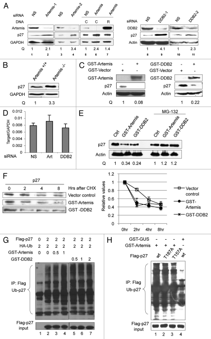
Artemis and DDB2 regulate p27 protein levels via a ubiquitin-mediated pathway. (A) P27 accumulates in Artemis or DDB2 depleted cells. HeLa cells were transfected with control (NS), Artemis or DDB2 siRNas. Forty-eight hours after transfection, cells were harvested and cell lysates were subjected to immunoblot analysis. Artemis-1 and Artemis-2, DDB2-1 and DDB2-2 indicate distinct siRNAs. As a control, 24 h after siRNA transfection cells were transfected with control plasmid DNA (C) or an Artemis construct refractory to Artemis siRNA (R). Cells were then incubated for an additional 24 h before harvesting. GAPDH and Actin indicate loading controls. “Q” indicates the relative band intensities of p27 normalized to GAPDH or actin levels. (B) p27 accumulates in Artemis null MEF cells. Lysates prepared from Artemis+/+ and Artemis−/− MEF cells were subjected to immunoblot analysis. (C) Overexpression of Artemis or DDB2 reduces p27 levels. HeLa cells were transfected with GST-Artemis (left part) or GST-DDB2 (right part) plasmid DNAs or treated with mock transfections. Forty-eight hours after transfection, cells were harvested and cell lysates were subjected to immunoblot analysis. (D) P27 mRNA is stable in Artemis or DDB2 depleted cells. mRNA was isolated from HeLa cells 48 h after transfection with control (NS), Artemis or DDB2 siRNAs. P27 mRNA levels were determined by real-time PCR. Results were normalized using GAPDH as an internal control. (E) Artemis and DDB2 regulate p27 protein levels through proteosome mediated degradation. HeLa cells were transfected with GST-Artemis or GST-DDB2 DNAs. After 48 h cells were treated with 20 mM MG-132, and 5 h later, cells were harvested and lysates subjected to immunoblot analysis. (F) Overexpression of either Artemis or DDB2 decreases the half-life of p27 compared with control. HeLa cells were transefected with GST-Artemis or GST control vectors. Thirty-six hours after transefection, cycloheximide (100 µg/ml) was added and the decrease in p27 protein level analyzed as a function of time by immunoblot analysis (left part). Quantitation of immunoblots are shown (right part). (G) Artemis and DDB2 promote ubiquitylation of p27 in vivo. HeLa cells were transfected with indicated amounts of Flag-p27, HA-Ub and GST-Artemis or GST-DDB2 DNAs. Cells were harvested 48 h after transfection, and an in vivo ubiquitination assay was performed using anti-Flag M2 agarose. Ubiquitylated proteins were detected using an HA antibody. Flag-p27 indicates a loading control at 10% of input. (H) Artemis promotes ubiquitylation of p27 independent of its phosphorylation on T187 or T157. HeLa cells were transfected with Flag-tagged wild-type (lane 1 and 4), T187A (lane 2) or T157A (lane3) p27, HA-Ub, GST-Artemis and GST-GUS. Forty-eight hours after transfection, an in vivo ubiquitylation assay was performed as indicated in Figure 4F.

