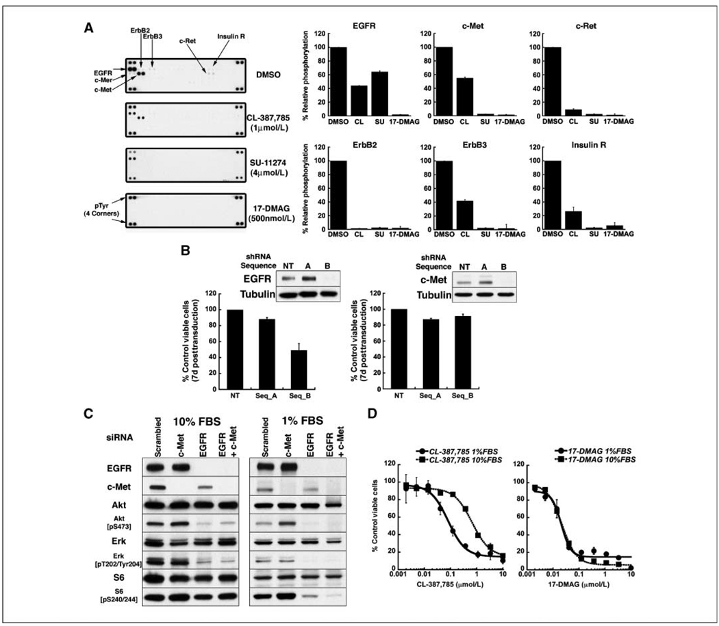
Figure 4.
NCI-H1975 (EGFR L858R/T790M) cells express activated RTKs in addition to mutant EGFR, which contribute to signal transduction and invasiveness and which are depleted by17-DMAG. A, left, lysates from NCI-H1975 cells treated with DMSO or the indicated compounds for 24 h were subjected to RTK profiling, as described in Materials and Methods, demonstrating the activation of multiple receptors in addition to EGFR. In each array, each RTK is assayed in duplicate. Right, densitometric analysis was performed and normalized for background and array lot variations. Relative phosphorylation of receptors in drug-treated cells was compared with that in DMSO-treated cells, the latter defined as 100%. Columns, average of duplicate spots; bars, SD. B, NCI-H1975 cells were transduced with lentiviruses encoding shRNAs targeting EGFR or c-Met. CCK-8 assays were performed 7 d after infection, and viability was normalized to cells infected with a lentivirus encoding a nontargeting shRNA construct. Western blots were performed 5 d posttransduction; in each case, the nontargeting construct and sequence A did not reduce expression of EGFR or c-Met. For shRNA targeting EGFR, columns represent the average of duplicate samples; for shRNA targeting c-Met, columns represent the average of quadruplicate samples. Bars, SD. C, NCI-H1975 cells were transfected with scrambled siRNA, or siRNA targeting EGFR or c-Met either alone or together. Transfected cells were maintained in growth medium with 10% or 1% fetal bovine serum; 48 h posttransfection, lysates were subjected to Western blotting with the indicated antibodies. Depletion of EGFR and c-Met in low serum is required to reduce mTOR signaling. D, NCI-H1975 cells were treated with the indicated concentrations of CL-387,785 or 17-DMAG in medium supplemented with 10% or 1% FBS. At 72 h, CCK-8 assaywas performed and the viability of each sample was normalized to that of DMSO-treated cells. Points, average of normalized values from two independent experiments; bars, SD. The IC50 for CL-387,785 in 10% serum is 667 nmol/L and decreases to 81 nmol/L in 1% serum. The IC50s for 17-DMAG in 1% and 10% serum were 21 and 22 nmol/L, respectively.

