
Fig. 1.
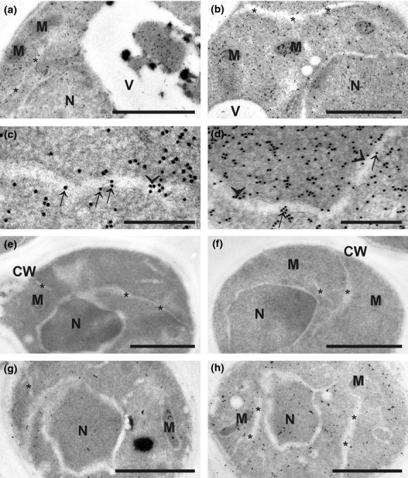
Transmission electron micrographs showing the subcellular distribution of glutathione in Saccharomyces cerevisiae grown on YPD (a, e–h) and SC−GSH media (b–d). Gold particles bound to glutathione could be found in mitochondria (M), nuclei (N), vacuoles (V), the ER (asterisks), and the cytosol of S. cerevisiae grown on YPD and SC−GSH media. Note that S. cerevisiae grown on SC−GSH medium (b) contain higher amounts of gold particles bound to glutathione in mitochondria than cells grown on YPD medium (a). Additionally, gold particles bound to glutathione could be observed in vacuoles of S. cerevisiae grown on YPD medium (a). Gold particles could also be found along the membranes (arrows in c) and also inside the lumen (arrowheads in c) of the ER in cells treated with 5 mM H2O2 for 60 min. Gold particles were also present in the perinuclear space (arrows in d) and along the inner membrane of the nucleus (arrowheads in d). No or only very few gold particles were detected in S. cerevisiae after sections were incubated with the primary antibody preadsorbed with an excess of oxidized glutathione (e) and with preimmune serum instead of the primary antibody (f). The glutathione-deficient mutants gsh1Δ (g) and gsh2Δ (h) contained markedly lower levels of gold particles when compared to the controls. CW, cell walls. Bars = 1 μm in a, b, e–h and 0.25 μm in c, d.